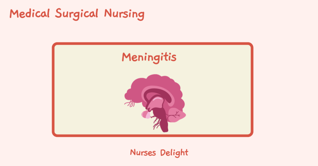
Meningitis is the inflammation of the meninges (dura mater, arachnoid and pia mater) that form protective layers over the brain and the spinal cord.
These layers have two main functions:
- They support the cranial and cerebral vasculature
- The protect the central nervous system from mechanical damage
The dura mater is the outermost layer and is located immediately beneath the cranium.
The arachnoid is the middle layer of the meninges. Located underneath is the subarachnoid space that contains the cerebrospinal fluid.
The pia mater is a thin layer that is located underneath the subarachnoid space and is direct contact with the surface of the brain and spinal cord. It follows the contours of the brain.
Causes of Meningitis
- Bacteria such as Streptococcus pneumoniae and Neisseria meningitidis.
- Viruses including enteroviruses, arbovirues, herpes simplex and mumps virus. Enteroviruses are the major cause of aseptic meningitis.
- Fungi. Cryptococcus neoformans, Coccidioides immitis and Blastomyces dermatitidis are the most common infections.
- Protozoa. Angiostrongylus contonensis and Gnathostoma spinigerum are the most renown parasites to cause meningitis.
- Cancer
- Weak immune system, for example in HIV patents
- Some drugs e.g., NSAIDS, antibiotics and intravenous immunoglobulins
Pathophysiology
Meningitis can occur in two ways:
Through the blood. This is usually referred to as hematogenous seeding. It occurs when bacteria in the bloodstream crosses the blood brain barrier to cause direct inflammation through immunity-mediated reactions.
Direct spread. Organisms enter the cerebrospinal fluid through infections of adjacent anatomic structures such as ear and sinuses (Otitis media and sinusitis respectively). It can also occur through injuries to the facial bones and invasive procedures.
Clinical Manifestations
- Headache
- Fever
- Neck immobility/ Nuchal rigidity. A stiff and painful neck can be an early sign.
- Positive Kernig sign. When the patient lies supine with the thigh flexed on the abdomen, the leg cannot be completely extended.
- Positive Brudzinski sign. When the patient’s neck is flexed, the knees and hips are also flexed. When one of the lower extremities is flexed, the opposite extremity exerts a similar movement.
- Photophobia
- Rashes, ranging from petechiae to large ecchymosis
- Disorientation and memory loss
- Seizures
- Lethargy and coma
- Increased intracranial pressure
Assessment and Diagnostic findings
- A CT scan is used to detect a shift in brain contents, risk of herniation, intracranial pressure and brain abscess.
- Lumber puncture is done to examine the cerebrospinal fluid.
- Bacterial culture and gram staining is done to isolate the causative agent.
- Complete blood count will reveal elevated levels of white blood cells, and in particular, polymorphonuclear leukocytosis with a left shift.
- Electrolytes to maintain fluid balance
- Chest, air sinuses and mastoids radiology. Chest x rays in patients with pneumococcal meningitis may reveal pneumonia.
Medical Management
- Early administration of antibiotics that cross the blood brain barrier in sufficient concentration to exert an efficacy e.g., Penicillin G in combination with Cephalosporins
- Amphotericin B and flucytosine may be used to treat fungal meningitis.
- Using Dexamethasone as adjunctive therapy in treatment of acute bacterial meningitis and pneumococcal meningitis
- Administer fluid volume expanders to treat dehydration and shock
- Administer anticonvulsant to treat seizure
Nursing Management
Assessment
- Neurologic and neurovascular status of the patient before initiation during therapy. Do cranial nerve assessment with attention to III, IV, VI, and VII.
- Assess the patients’ vitals.
- Perform ABG and pulse oximetry tests to identify need for respiratory support
- Assess for complications like increased intracranial pressure and hydrocephalus
Nursing Diagnosis
- Hyperthermia related to meningeal infection as evidenced by high body temperature
- Ineffective tissue perfusion related to cerebral edema as evidenced by high carbon IV oxide in the blood (hypercapnia)
- Acute pain related to increased intracranial pressure as evidenced by headache
- Ineffective airway clearance related to neuromuscular damage as evidenced by dyspnea
- Risk for injury related to decreased loss of consciousness and altered sensorium
Nursing Interventions
- Ensure proper infection control standards are met.
- Pain management with relevant pain medications
- Antipyretics and cooling blankets to treat elevated temperatures
- Encouraging hydration either orally or peripherally
- Neurologic and vitals monitoring and every 2 to 4 hours
- Protecting the patient from injury secondary to seizure activity or altered LOC
- Monitor daily weight serum electrolytes and urine volume, specific gravity, input and output
- Prevent complications related with immobility e.g., pressure ulcers and
- prioritize care to maintain airway, breathing and circulation
- Provide a quiet environment. Minimize bright lights to windows and overhead lights Maintain bedrest with head elevated 30 degrees
- Monitor and prevent complications; Increased intracranial pressure, vascular dysfunction, fluid and electrolyte imbalance, seizures shock.


